News|Articles|March 5, 2026
Hi-C Mapping Reveals Hidden "Enhancer-Hijacking" in Lymphoid Malignancies
Author(s)Paige Britt
Fact checked by: Andrea Eleazar, MHS
Listen
0:00 / 0:00
Key Takeaways
- Hi-C provides genome-wide partner identification for SVs from FFPE tissue, surpassing locus-limited FISH and complementing WGS by directly inferring functional cis interactions between distal enhancers and promoters.
- Enhancer hijacking emerges as a major “missing driver” mechanism in LBCL/PCN, enabling detection of non-chimeric oncogene activation such as IGL::BCL2, IGH::CCND2, and MAFA/MAFB rearrangements.
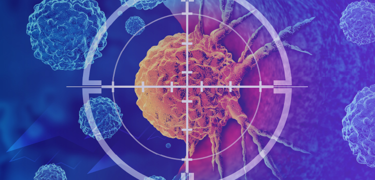
Hi-C on routine FFPE biopsies uncovers hidden lymphoma drivers, enhancer hijacking, and PD-L1 rearrangements, guiding targeted therapy beyond FISH.
The diagnostic landscape for lymphoid cancers—spanning large B-cell lymphomas (LBCL), plasma cell neoplasms (PCN), and acute lymphoblastic leukemias (ALL)—is currently tethered to decades-old technologies. While fluorescence in situ hybridization (FISH) remains the gold standard for detecting structural variants (SVs), its low-throughput nature and "blind spots" for variant rearrangements often leave clinicians without a clear driver for their patients' disease.
A pivotal study in Cell Genomics has now demonstrated that Hi-C, a chromosome conformation capture method, can be successfully applied to routinely processed formalin-fixed, paraffin-embedded (FFPE) clinical biopsies.1 By mapping the 3-dimensional architecture of the genome, Hi-C not only identifies fusions missed by FISH but also characterizes the "enhancer-hijacking" events that drive oncogene overexpression.
Overcoming the Limitations of Standard Cytogenetics
Current clinical workflows for FFPE samples face significant technical hurdles. FISH is restricted to a handful of loci and often fails to identify partner chromosomes. Meanwhile, whole-genome sequencing (WGS) struggles with repetitive regions and cannot confirm the functional cis interactions between distal enhancers and promoters.
In contrast, Hi-C maps pairwise topological interactions, providing a "topological blueprint" of the tumor. The study confirmed that Hi-C is robust enough for clinical use, yielding high-quality data from archival samples up to 17 years old.
Key Advantages of Hi-C in the Clinic:
- Comprehensive detection: Identifies partners for all rearrangements genome-wide in a single assay.
- Structural resolution: Distinguishes between functional drivers and "passenger" events.
- Copy number insights: Detects critical alterations, such as CDKN2A/B loss and whole-chromosome gains.
Identifying the "Missing" Drivers: Enhancer Hijacking
One of the study's most significant contributions is the detection of enhancer-hijacking. This occurs when a rearrangement places an oncogene adjacent to a distal enhancer, driving massive expression without creating a chimeric protein.
In several cases, Hi-C identified clinically relevant drivers that were completely missed by standard FISH panels:
- DLBCL: An IGL::BCL2 rearrangement was identified where standard probes failed.
- Multiple myeloma: An IGH::CCND2 rearrangement—a rare but aggressive driver—was detected.
- MAF family: Rare MAFA and MAFB rearrangements, often excluded from routine testing, were functionally validated through Hi-C topological data.
Hi-C revealed unanticipated rearrangements involving programmed death-ligand genes. In cases of primary central nervous system lymphoma and primary mediastinal B-cell lymphoma, rearrangements juxtaposed CD274 (PD-L1) or PDCD1LG2 (PD-L2) with potent enhancers. This finding provides a mechanistic rationale for using checkpoint inhibitors like pembrolizumab (Keytruda) in refractory cases.
Hi-C allows clinicians to move beyond simple "break-apart" results to understand the functional consequence of a rearrangement.
BCL6: Driver vs Passenger
Standard FISH might show a BCL6 break, but Hi-C can categorize the event:
- Promoter-replacement: Drives BCL6 expression (gene-activating).
- Enhancer-donating: A "pseudo-double-hit" where BCL6 enhancers are donated to activate MYC.
- Passenger events: Complex variants with no evidence of oncogenic interaction.
The research highlighted that the MYC locus possesses distinct "interaction domains" tailored to the specific cancer type. This "cell-state-selective" topology dictates where functional breakpoints are likely to occur.
Implementation Challenges
While the study proves Hi-C's utility, hurdles to widespread clinical adoption remain. The method requires significant sequencing depth (averaging approximately 260 million read pairs per sample), leading to higher costs. Additionally, the current 25kb resolution may miss small focal deletions, and subclonal events (present in <15% of cells) remain difficult to detect.
As precision oncology moves toward functional genomics, Hi-C offers a sophisticated lens through which to view the "rewired" genomes of lymphoid cancers. By identifying fusions and enhancer-hijacking events that escape traditional testing, Hi-C may soon provide the definitive structural map required for personalized treatment strategies.
“This study from our collaborators at the University of Michigan and [New York University] provides further evidence of how the whole-genome approach enabled by Hi-C sequencing addresses the limitations of conventional methods for the detection of rearrangements that drive classification and care decisions in lymphoid cancers,” said Anthony Schmitt, PhD, senior vice president of Science at Arima Genomics, in a news release.2
REFERENCES
1.Wu J, Chu SC, Cho J, et al. Hi-C for genome-wide detection of enhancer-hijacking rearrangements in routine lymphoid cancer biopsies. Cell Genomics, Volume 0, Issue 0, 101166. Published February 20, 2026. Accessed March 3, 2026.
2.Arima Genomics announces publication of new study supporting the whole-genome rearrangement detection approach behind Aventa Lymphoma. News release. Arima Genomics. Published February 26, 2026. Accessed March 3, 2026. https://tinyurl.com/yc5fc7jc
Newsletter
SubscribeRelated Content
Advertisement